The Grand Symphony of Erection: A Journey Through Penile Anatomy and Function
In the vast tapestry of human biology, few mechanisms are as exquisitely orchestrated, as profoundly complex, and yet as elegantly simple in their outcome as the erectile process. It is a testament to millions of years of evolutionary refinement, a marvel of hydraulic engineering, neurochemical precision, and vascular ingenuity, all working in concert to achieve a singular, vital purpose. For the knowledgeable mind, understanding the anatomy of erectile function is not merely a dry recitation of parts and labels, but an immersive journey into a living symphony, where every cell, every nerve ending, every molecule plays a critical, indispensable role.
Let us embark on this journey, not as detached observers, but as privileged witnesses to a biological ballet, a story of intricate design unfolding within the very core of human experience.
Chapter 1: The Penile Pillars – Corpora Cavernosa and Their Unyielding Embrace
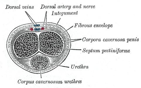
Our story begins with the fundamental structures, the very architecture that defines the potential for tumescence. At the heart of the penis lie three cylindrical columns of erectile tissue. Two of these, the corpora cavernosa (singular: corpus cavernosum), are the principal protagonists of erection. Imagine them as two parallel, sponge-like tubes, running the length of the penile shaft, from their anchors (crura) to the base of the glans.
From a macroscopic perspective, the corpora cavernosa appear robust, yet their true marvel lies within. Microscopically, they are a labyrinthine network of interconnected vascular spaces, or sinusoids, separated by delicate walls of smooth muscle and connective tissue called trabeculae. Think of these trabeculae as an intricate scaffolding, providing structural integrity while simultaneously housing the very smooth muscle cells that will dictate the flow of blood. These sinusoids, in their flaccid state, are largely collapsed, allowing only a trickle of blood to pass through. They patiently await their command.
Encasing each corpus cavernosum, and indeed the entire penile shaft, is a crucial layer of dense, inelastic connective tissue known as the tunica albuginea. This fibrous sheath is not merely a protective wrapper; it is the ultimate determinant of penile rigidity. Picture it as a taut, unyielding velvet glove, strong and resilient. In the flaccid state, it maintains the shape of the penis. But during erection, its inelasticity becomes paramount. As blood rushes into the cavernous spaces, the tunica albuginea stretches to its limit, like a balloon inflating inside a rigid casing. This stretching is key to compressing the delicate venules that drain blood from the corpora, effectively trapping the blood within and creating the hydraulic rigidity characteristic of a full erection. Without a robust tunica albuginea, the penis would swell but never achieve true firmness, a condition akin to a leaking tire. Its fibrous collagen and elastic fibers are meticulously arranged, forming an almost impenetrable barrier that transforms the soft tissue into a rigid column.
The two corpora cavernosa are separated internally by a fibrous septum, which, though mostly complete, has fenestrations (small openings) allowing blood to flow freely between them, ensuring a unified hydraulic system. This interconnectedness means that an erection is a holistic event for both corpora.
Chapter 2: The Urethral Guardian – The Corpus Spongiosum
While the corpora cavernosa are the engines of rigidity, the third column of erectile tissue, the corpus spongiosum, plays an equally vital, albeit different, role. This single column lies ventral (below) to the corpora cavernosa, completely encircling the urethra, the conduit for urine and semen. Unlike its cavernous counterparts, the corpus spongiosum is designed for a less intense, yet critical, degree of engorgement.
Its primary function during erection is to prevent the urethra from being compressed by the expanding corpora cavernosa. Imagine the hydraulic pressure building in the corpora; without the protective, albeit lesser, engorgement of the corpus spongiosum, the urethra would be pinched shut, making ejaculation impossible and potentially causing discomfort. The corpus spongiosum, therefore, swells sufficiently to keep the urethral lumen patent, ensuring that the pathway remains open.
At its distal end, the corpus spongiosum expands to form the glans penis, the highly sensitive tip of the penis. While the glans also becomes engorged during erection, it does so to a lesser degree than the corpora cavernosa, maintaining its characteristic texture and sensitivity. Proximally, the corpus spongiosum expands to form the bulb of the penis, which is anchored to the perineum. This continuity underscores its role in the entire erectile complex.
The venous drainage of the corpus spongiosum is also distinct, featuring a more robust outflow system that prevents it from becoming as rigid as the corpora cavernosa. This differential rigidity is a masterpiece of design, allowing the penis to be firm enough for penetration while remaining sufficiently supple at its tip to maximize sensation and protect the urethra.
Chapter 3: The Vascular Network – The Lifeblood of Erection
No story of hydraulic function is complete without its fluid dynamics, and in the penis, this is a tale told in arteries and veins. The vascular system is the plumbing, the intricate network that brings the necessary resources and removes the byproducts, all under precise neural and chemical control.
The Arterial Supply: The Inflow Architects
The journey of blood into the penis begins with the internal pudendal artery, a major branch of the internal iliac artery. As it progresses, it gives rise to three crucial penile arteries:
-
The Cavernosal (Deep) Arteries: These are the primary vessels feeding the corpora cavernosa. Each corpus cavernosum receives its own deep artery, which runs longitudinally through the center of the erectile tissue. These arteries are not straight pipes; they branch repeatedly, giving rise to numerous smaller arterioles. The most critical of these smaller vessels are the helicine arteries. These are coiled, highly muscular arteries that, in the flaccid state, maintain a high basal tone, restricting blood flow into the sinusoidal spaces. They are the gatekeepers, the precise valves that, upon receiving the command for erection, will relax and open wide, unleashing a torrent of blood. Their unique helical shape allows them to straighten and dilate dramatically, maximizing flow.
-
The Dorsal Arteries: These two arteries run along the dorsal (top) surface of the penis, just beneath the tunica albuginea. They supply blood to the tunica albuginea itself, the skin of the penis, the glans, and the corpus spongiosum, contributing to the overall engorgement and health of these structures.
-
The Bulbar Arteries: These arteries supply the bulb of the penis and the posterior portion of the corpus spongiosum, reinforcing its role as the urethral guardian.
The coordinated dilation of these arterial systems, particularly the helicine arteries, is the initial spark of erection, the moment the floodgates open.
The Venous Drainage: The Outflow Regulators


Post Comment